


Pathology Course
July 1 - 4, 2025
Amsterdam, The Netherlands
June 24 - 27, 2025
Basel, Switzerland
- specialize in nephropathology
- 3 months fellowships supported by the European Society of Pathology
- advanced training in nephropathology (or other specialized fields)
- for young ESP-members at the beginning of their careers
- next application deadline: June 2025

Data Base for Chemicals, Drugs, Poisons and other related Medical Interventions
Our Mission
The mission of our working group is the advancement of nephropathology, including transplant pathology in Europe. The working group supports continuous education and training in renal pathology. It promotes research, organizes collaborative studies, and offers help in diagnostic cases.
Who We Are
We are pathologists with a special training and expertise in nephropathology. We are organized as a working group within the European Society of Patholgy (ESP). The working group is run by an elected chair and co-chair as well as all active members that want to be involved. Read more …
What We Do
Together, we organize the nephropathology content of the annual European Congresses of Pathology (read more …). Our members are involved in educational activites and scientific meetings within Europe and worldwide. We provide technical information about the work-up of native and transplant kidney biopsies. We train and mentor young pathologists in our field to achieve the best diagnostic service for our clinical colleagues. Our group represents the European Society of Pathology(ESP) in affiliated learned societies and congresses and serves as knowledge provider to the ESP in the field of nephropathology.
Membership
The working group is open to all members of the European Society of Pathology at no additional cost. To become an ESP member, please visit the ESP website. To apply for the working group membership, use the membership login on the ESP website, go to profile, and press the “membership in working group” button.
History
The Nephropathology Working Group was founded at the occasion of the 19th Congress of the ESP on the initiative of Dusan Ferluga (Slovenia) in 2003. Past chairmen were Dusan Ferluga, Slovenia (2003-2007), Michael J. Mihatsch, Switzerland (2007-2013), Ian S. Roberts, United Kingdom (2013-2016), and Kerstin Amann, Germany (2016-2021).
Information for Laymen
Pathology is the study and diagnosis of disease through examination of organs, tissues, cells, and body fluids. The term encompasses both the medical specialty which uses tissues and body fluids to obtain clinically useful information, as well as the related scientific study of disease processes. Anatomical pathologists diagnose disease and gain other clinically significant information through the gross and microscopic visual examination of tissues, with special stains and immunohistochemistry employed to visualize specific proteins and other substances in and around cells, and electron microscopy to visualize ultrastructural changes. More recently, molecular biology techniques are also included in the study of tissues to gain additional clinical information. Anatomic pathologists serve as the definitive diagnosticians for most cancers, as well as numerous other diseases. Nephropathologists are specialized anatomical pathologists. They have special expertise for the study of renal diseases in native and transplanted kidneys.
A collaborative scientific project by Amélie Dendooven, Sabine Leh, and Mark Helbert.
Revisiting proliferative glomerulonephritis with monoclonal immunoglobulin deposits through immunoglobulin repertoire sequencing
By:
[1] Javaugue V, Pascal V, Nasr SH, et al. Revisiting proliferative glomerulonephritis with monoclonal immunoglobulin deposits through immunoglobulin repertoire sequencing. Kidney Int. 2025;108(6):1146-1157. doi:10.1016/j.kint.2025.07.039
Blogged by Nicolas Kozakowski, January 2nd, 2026
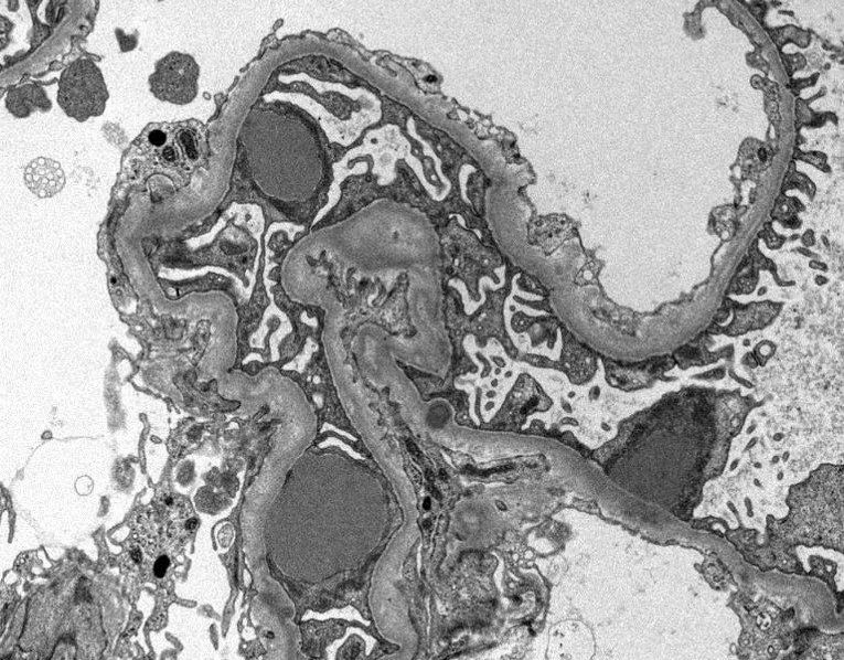
Proliferative glomerulonephritis with monoclonal immunoglobulin deposits (PGNMID), originally described as a proliferative GN with monotypic IgG deposits, most often IgG3, is now classified as a monoclonal gammopathy of renal significance–associated disease. Given its rarity, renal pathologists consider PGNMID a diagnostic trophy. Yet, the true prey often remains elusive, as a circulating monoclonal immunoglobulin or the causative clone is rarely captured.
Using a highly sensitive high-throughput sequencing assay for immunoglobulin mRNA, investigators recently embarked on a systematic clone hunt in a multicentre PGNMID cohort, supplemented by proteomics and immunofluorescence. Their expedition uncovered an attributable clonal proliferation matching the biopsy findings in roughly one quarter of cases, while the majority yielded no such quarry. Further tracking using antibodies against light-chain variable domains revealed that clone-positive cases showed variable-domain restriction, whereas clone-negative cases displayed a heterogeneous pattern, suggesting oligoclonal deposits.
These findings imply that many cases currently labelled PGNMID may represent antigen-driven oligoclonal immune responses rather than true monoclonal gammopathies and may warrant reclassification.